For decades, transplant medicine has replaced kidneys, livers, hearts, lungs, hands, and faces. The eye remained different. Its delicate structures are connected to the brain by the optic nerve, a bundle of millions of fibers that medicine has not yet learned to reliably repair.
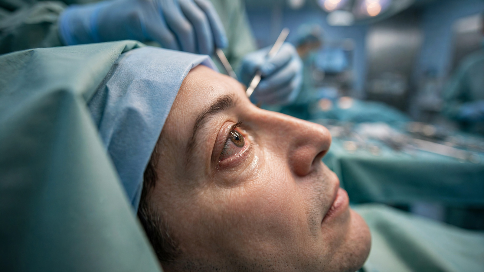
That is why a procedure carried out in New York in 2023 became such an important moment in modern surgery. A team at NYU Langone Health transplanted an entire human eye into a living patient as part of a partial face transplant. The eye did not restore the patient’s sight, and doctors have not solved the problem of reconnecting it to the brain. But the transplanted organ survived, retained blood flow, and remained healthy during follow-up.
The result was not a finished treatment. It was something more useful at this stage: evidence that a whole human eye can be moved from one body to another and kept alive.
A different kind of transplant challenge
Corneal transplantation has been performed for more than a century. In that procedure, surgeons replace the transparent front layer of the eye, often improving vision for people whose corneas have been damaged by disease or injury.
A whole-eye transplant is far more demanding. The operation involves the eyeball itself, the retina at the back of the eye, the blood vessels that nourish it, and the optic nerve that carries visual information toward the brain. Each part must be protected during removal and connected to the recipient’s anatomy with extraordinary precision.
The optic nerve presents the largest biological obstacle. When it is cut, the nerve fibers generally do not grow back across the gap in a way that restores normal vision. Surgeons can reconnect blood vessels. They can align muscles and repair skin. They can place the eye in the orbit. They cannot yet recreate the original network linking the eye to the visual centers of the brain. For related reading, see Sesame Street: How a Neighborhood on Television Made Learning Feel Like Belonging.
That limitation shaped the meaning of the NYU Langone operation. The immediate goal was not to promise sight. It was to test whether the eye could remain structurally and biologically viable after transplantation—and whether the procedure could be safely combined with reconstruction of the face.
The injury that led to the operation
The patient, Aaron James, was a utility worker from Arkansas who suffered a devastating electrical accident in 2021. The injury severely damaged the left side of his face and destroyed his left eye. He also faced the loss of facial structures needed for eating, breathing, speaking, and appearance.
Traditional reconstruction could repair some of the damage, but it could not replace everything that had been lost. A donated face offered the possibility of restoring a more complete set of structures at once. The medical team decided that the same donor could also provide an entire eye.
In May 2023, surgeons performed a procedure that lasted about 21 hours. The operation involved more than 140 medical professionals, according to NYU Langone. It combined a partial face transplant with transplantation of the left eye and its surrounding tissues.
The face transplant included structures such as the nose, lips, cheek, and eyelids. The eye transplant required surgeons to work around the orbit, connect major blood vessels, and position the eye within the recipient’s socket. The team also used adult stem cells obtained from the donor’s bone marrow in an effort to support the optic nerve and the transplanted tissues.
That stem-cell approach was experimental. It was not expected to instantly regenerate the optic nerve, but it reflected a broader strategy: if doctors cannot yet rebuild the entire nerve pathway, they may still be able to improve the environment around the nerve and preserve the eye while future therapies develop.
What doctors saw after surgery
The first test was survival. Transplanted organs can fail when blood supply is inadequate, when the immune system attacks them, or when infection and other complications overwhelm the patient. The eye passed several important early tests. For related reading, see The Human Pangenome: How Scientists Are Building a More Complete Map of Our DNA.
Doctors reported that the eye had good blood flow through the retinal artery and vein. The retina remained viable, and the pressure inside the eye stayed within a healthy range during the reported follow-up. The patient also avoided rejection of the transplanted eye during that period.
Those findings mattered because the eye is not simply a camera that can be placed into an empty socket. It is living tissue that depends on a continuous supply of oxygen and nutrients. A successful transplant therefore requires the eye to become part of the recipient’s circulation, not merely to appear anatomically complete.
At the same time, the operation did not produce useful vision. The transplanted eye could not send normal visual signals to the brain because the optic nerve had been severed. The patient continued to see with his remaining eye.
That distinction is essential. Describing the procedure as a restored-sight breakthrough would go beyond the evidence. Its achievement was more fundamental: it showed that the biological life of a whole eye can be preserved after transplantation, even when sight remains out of reach.
Why the result still changes the field
Medical progress often begins with a procedure that answers one question while leaving the hardest question unresolved. In this case, surgeons learned that they could transplant the eye and maintain its blood supply. That creates a platform for research into the next problem: reconnecting the eye to the nervous system.
Future work could involve therapies designed to encourage optic-nerve fibers to regenerate, guide them toward the brain, and restore the precise connections needed for vision. Those are separate challenges, and solving one would not automatically solve the others. Researchers must also understand how the visual system would interpret signals from a transplanted eye after a long period of blindness. For related reading, see Arizona’s Apache Trout Recovery Shows What Patient Conservation Can Do.
The operation may also help people who suffer severe facial injuries even when vision cannot be restored. A functioning transplanted eye can help preserve the shape of the face, support eyelid reconstruction, and provide a more natural physical result. For patients with major trauma, those outcomes can affect comfort, protection of the remaining eye, speech, eating, and social life.
Face and eye transplantation also raise difficult questions. Recipients generally need lifelong immunosuppressive medicines to reduce the risk of rejection. Those drugs can increase vulnerability to infection, kidney problems, and other complications. Because these operations are rare and highly specialized, every decision must weigh potential benefits against substantial medical risks.
A beginning rather than a final victory
The first whole-eye transplant was not a miracle cure, and it did not make blindness reversible. It was a carefully monitored experiment that succeeded in a narrower but important way: a human eye from one person became living tissue in another person.
That achievement moved the boundary of transplant surgery. It demonstrated that the eye’s blood supply could be restored, that the organ could remain healthy, and that whole-eye transplantation could be performed alongside complex facial reconstruction.
The next breakthrough may come from regenerative medicine rather than surgery alone. Scientists are studying nerve growth, stem cells, bioengineered tissues, and methods for repairing the connection between the eye and brain. None has yet produced a routine treatment that restores sight after a severed optic nerve.
But the NYU Langone procedure gives those efforts something they did not previously have: a living, transplanted human eye that can be observed over time. The operation did not complete the journey to restored vision. It established that the journey has a place to begin.
Use: Primary institutional account of the operation, surgical team, patient outcome, eye viability, and follow-up.
Use: Peer-reviewed clinical report describing the transplant procedure and postoperative findings.
Use: Authoritative background on the optic nerve and how visual information travels from the eye to the brain.